
With the growing area of regenerative medicine, it’s no surprise that there are dozens of systems available to process Platelet Rich Plasma (PRP). While the use of each system will result in a plasma product, the processing techniques, equipment and final sample differ quite drastically. In this installment of our Myth Buster series, we will take a closer look at the differences that one can expect to encounter when looking at PRP processing systems and how each of these components effects the PRP product.

Processing Techniques
There are three main methods by which Platelet Rich Plasma can be processed. These methods are:
1. Centrifugation – This is the most common technique available, which involves using a centrifuge to separate the cell layers in whole blood or to pass the blood components through a gel separator (which is in the device prior to insertion of blood) to isolate the platelets from other cell types.
 a. Spinning without gel separator – When blood is spun at a high rate of speed for a period of time, the cellular components will separate according to their density within the container/tube. These cell layers can be visualized upon removal of the device as seen in the below picture. The three distinct layers of blood include the plasma layer, the platelet buffy coat and the red blood cell layer. The plasma and buffy coat can be collected and injected or the suspension can be placed in a second container for a second spin. The second spin in the process enables further concentration of the platelets, which as we discussed in our previous posts, is important for delivering a therapeutic concentration of platelets (between 3-7-fold concentration compared to circulating blood). A sample that is obtained by a single spin process will have a lower concentration of platelets, thus possibly resulting in a sub-optimal therapy.
b. Spinning with gel separator – Gel separators provide a physical gradient for blood to pass through resulting with the final product at the top of the tubes. Gel separators are typically used for isolating either plasma or serum, but have been developed to allow smaller cell types to remain in the final product. Unfortunately, the majority of products available that use gel separation produce a low concentration of platelets due to the small volume of blood being used (typically 8- 10 mL) as well as the poor capture of platelets in the final product.
2. Flow Cytometry - This method involves a highly specialized piece of equipment that utilizes the absorption of light by a cell and further separates the cells according to the refracting light. This process relies on blood flowing in a single layer suspension through a complex network of tubes, which can be further complicated if the sample begins to clot during the process. Flow cytometry is typically automated, which provides great convenience to the user, but the equipment may require additional set up (prior to processing) and upkeep to ensure proper calibration for processing samples.
3. Gravity and Reverse Osmosis – This method involves utilizing a specially designed blood containment system with a built-in filter. The filter’s purpose is to collect the platelets while allowing the other cells to pass through. Although this method is attractive from a space and equipment perspective, performance of these systems has shown to result in an increase of neutrophils which can be detrimental when injected into a joint.
Volume of Blood
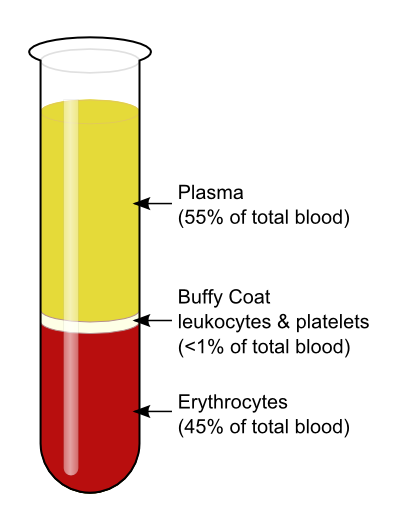 One of the biggest differences between PRP processing systems is the volume of blood that is required to obtain the PRP. Some systems require as low as 8 mL of whole blood, while other systems, such as the CRT system, require between 25-50 mL. What does this variation mean to you? If we look at typical blood composition, as depicted in the picture below, we will see that platelets make up less than 1% of the circulating blood. In order to have a therapeutic dose of platelets in the volume which is necessary for administration, a higher volume of blood is required for processing. For example, when using the CRT system, it is recommended to collect 50 mL of whole blood to produce 4-5 mL of PRP (with concentration of platelets between 3-7 fold). For systems that require the smaller whole blood volume with the same end product volume, the concentration of platelets will be below the recommended therapeutic threshold.
Sterility/ Exposure to Environment
When considering any product that will be administered intra-articularly or into areas of poor blood flow, it is imperative to ensure that the processing is done with aseptic technique. It is also important that the exposure of the sample to environmental factors is minimal to reduce the likelihood of contamination. The majority of systems available are considered to be closed systems in which the sample being processed has little to no contact with the outside environment. There are some processing systems available which utilize open tubes or require passing the blood through a needle into the containment device, which may introduce additional contaminants into the sample.
Time to Process- From Collection to Administration
Depending on the technique of isolating the platelets (centrifuge vs. flow cytometry vs. gravity filter), processing times vary greatly from just under 10 minutes to as much as 45 minutes! Some processing systems require activation of the platelets, which can take up to 45 minutes. Activating platelets, however, is not a necessary step, since the platelets will activate and release their stored growth factors once they are exposed to collagen in the joint/tissue. The Companion Regenerative Therapies System takes less than 15 minutes from blood collection to administration, making it a fast, in-house therapy that can be easily scheduled even in a busy hospital.
To learn more about Platelet Rich Plasma and how it works as a therapy, watch this short animation:
Stay tuned for our next blog post where we “bust” another regenerative medicine myth!

