
 Next to its unique characteristics allowing patient stress, fear, and pain to be reduced, photobiomodulation therapy (PBMT) also has the ability to be utilized as an adjunct to standard of care for a myriad of conditions affecting species ranging from the smallest avian to the biggest elephant. PBMT offers us not only a noninvasive and painless tool, but it also affords the operator certain flexibility in its applications, as per the level of tissue disruption involved.
Certainly, each case is an individual instance and must be approached with an individual treatment design and delivery plan. Just as no two patients are the same, neither are the specifics surrounding the etiology / progression / current status and treatment care / etc. of each case. PBMT offers us the flexibility of addressing cases where there is any level of pain, inflammation, and/or tissue disruption. Most, if not all the patients we see, regardless of species, are likely to fit the bill in at least one of the three categories cited. Let’s take a look at some of the commonly-used ways in which a clinic can implement a successful PBMT program as an adjunct to standard of care.
Typically, a new adopter the modality will begin by using it for cases responsive to a short treatment course. Usually, these include acute conditions involving superficial tissues, such as pyotraumatic dermatitis and post-operative incisions. Naturally, as the operator’s knowledge base grows, so will the daily applications of the modality. Most operators report a positive experience in delivering this level of care. It is as therapeutic for the operator to provide this leading-edge level of care as it is for our patients to undergo it. The most challenging part of starting a successful and multifaceted PBMT program in a clinical setting is to overcome inertia. Once the ball gets rolling, it will naturally pick up speed.
 Once the operators have applied the early stages of making PBMT a core part of their multimodal approach to pain management, then the next natural evolution is to incorporate it with routine anesthetic procedures. Here, we see patients undergoing either a surgical or dental health procedure. In this setting, the therapy laser platform can again be utilized in a series of ways, from conditions such as gingivitis (the only reversible dental disease), going all the way up to multiple extraction sites or stomatitis. With surgical procedures somewhat more involved, like an extracapsular cruciate repair, a pre-op and post-op treatment would also prove to be of value. PBMT should also be highly considered as part of a convalescent care plan, especially when dealing with invasive or orthopaedic procedures (e.g. – FHO, TPO, TPLO, TTA, limb amputation, etc).
As with anesthetic patients, hospitalized patients should be given specific consideration and be offered the benefits of PBMT, especially while they are on location. Such examples that have shown the value of PBMT to standard of care include pancreatitis, HBC, degloving injuries, and snakebites, just to name a few. Typically speaking, “time is tissue” when it comes to injury to tissues (both soft and dense). The savvy laser operator is able to embrace this concept and thus understands to have a certain window of fluidity in the application of the modality. The operators’ knowledge base in PBMT and ability to be flexible with an in-patient approach treatment design and delivery, enables them to best address the individual needs and caveats of each case as a separate application (i.e. special considerations such as: active hemorrhage, neoplasia, or active growth plates).
Once a practice has reached this level of understanding and has applied a level of commitment in incorporating PBMT with their core values and message, and confident in its application, the final step is to incorporate it with long-term care plans. In this setting, the focus is on outpatient appointments, scheduled much as they would be for a DVM seeing outpatients. These are the long-term patients with incurable conditions where our goal (and reasonable expectation) is to manage the condition and prevent an active decline. Often, once a clinic gets to this level of focused care, a specific “daily designated laser operator” is usually assigned to the task of handling the daily appointments (larger practices with a sizeable technical staff will incorporate a rotation of daily operators so as to have everyone remain proficient in their technique).
Most practices successful in their integration and implementation of PBMT within their departmental daily modus operandi, in addition to the current standard of care, have proven the modality a synergistic behemoth in our ability to continue to adapt to, adopt, and successfully implement a dynamically evolving aspect of veterinary medicine. The successful and practical applications of PBMT are limited only by the specifics of the case and the ability of an operator to perceive the modality’s application for a specific case presentation. The initial inertia previously mentioned is quickly overcome and replaced by a momentum which will help propel any practice to the next level of patient care when PBMT is allowed to fully develop as a medical modality in clinical practice.
When utilizing therapy laser units, ocular protection is the primary safety concern. Exposure to the eye is at the top of a short list of contraindications for photobiomodulation therapy (PBMT). While this medical modality is not applicable to this physical structure, it is of great value for a myriad of conditions affecting other parts of the body. We need to take precautions to prevent accidental ocular exposure, and we would never intentionally laser the eye itself. That being said, an experienced operator can certainly do periorbital treatments as long as they protect the eye itself.

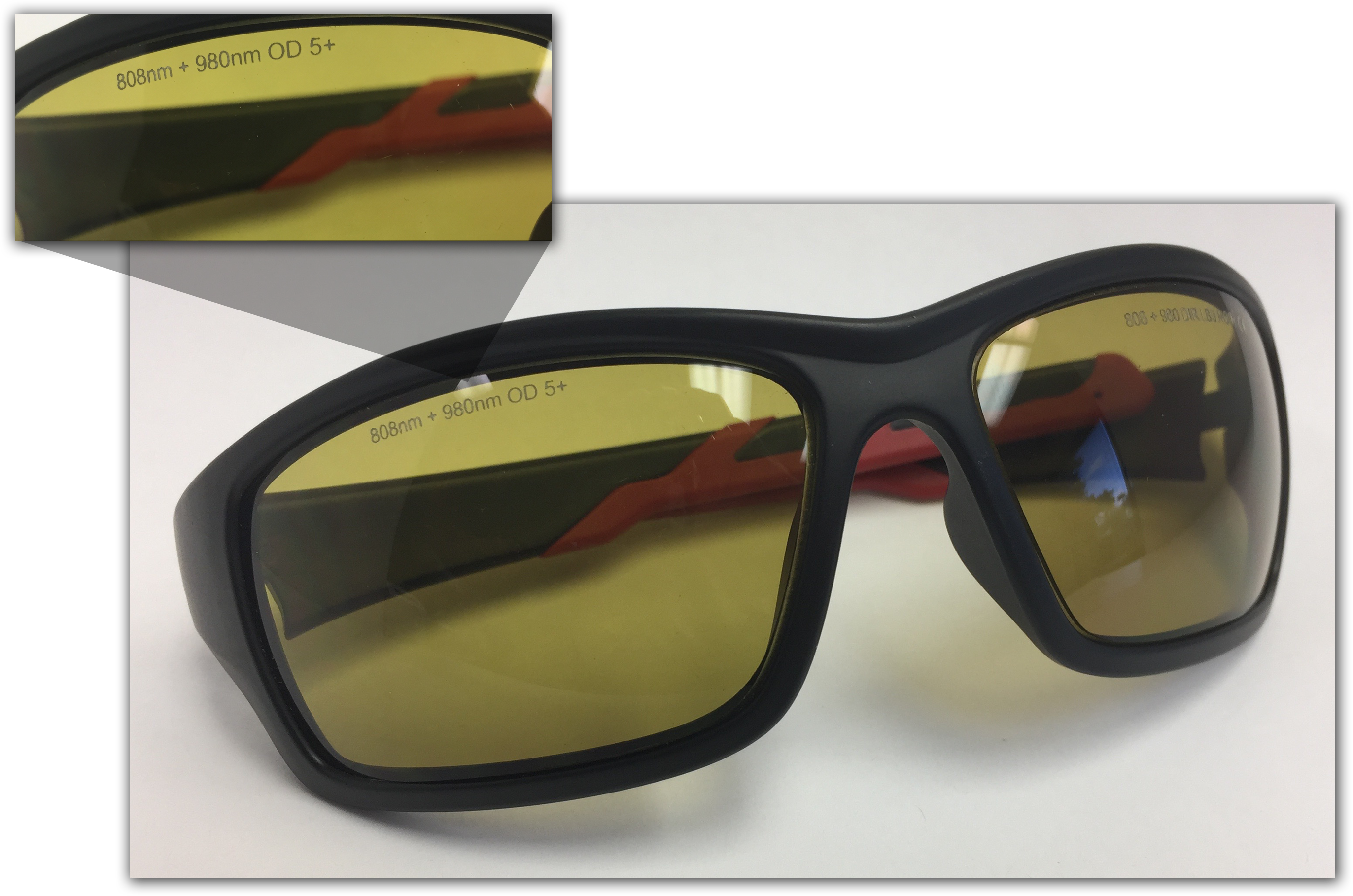
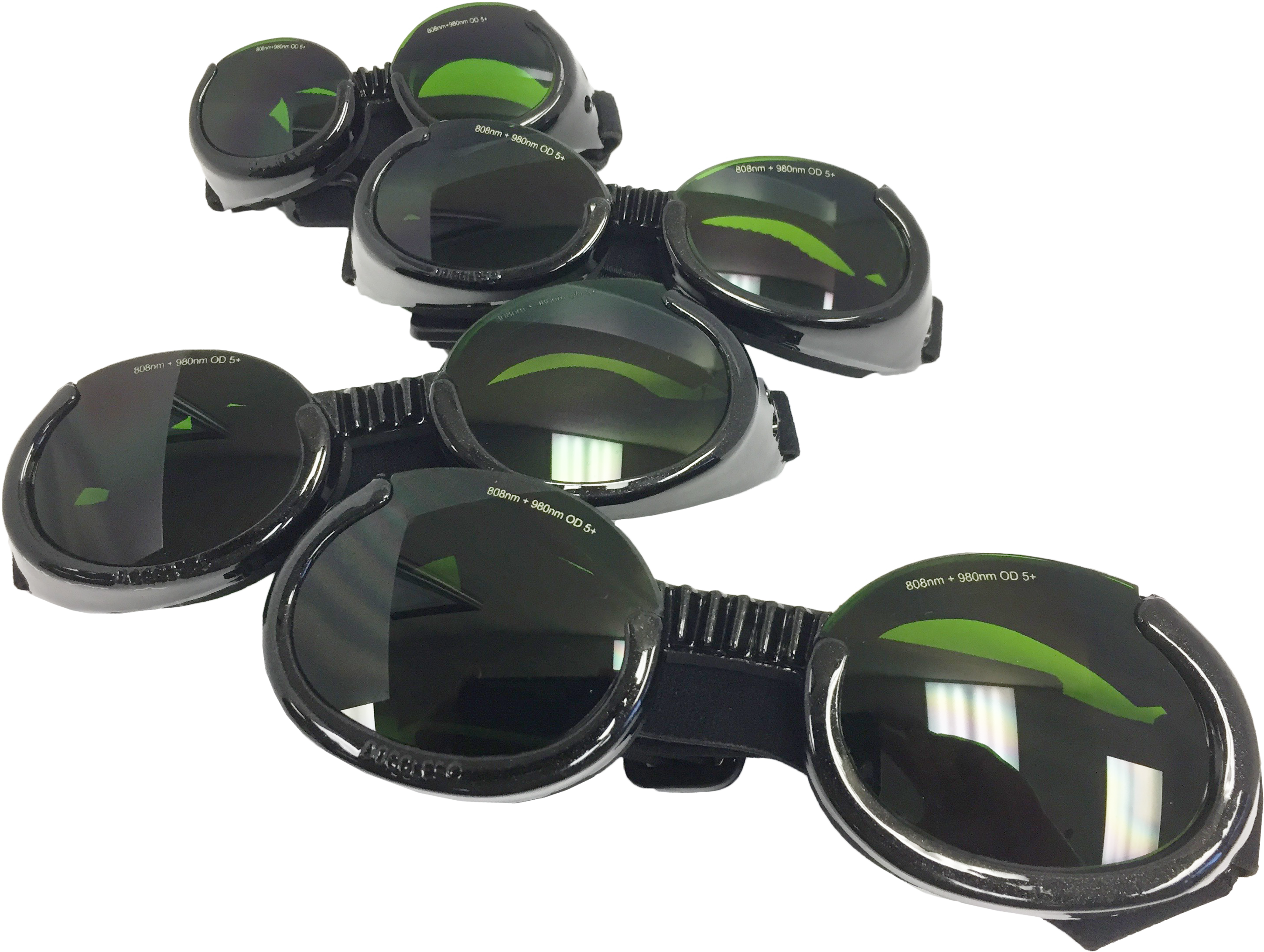
Appropriate ocular protection is easily acquired and achieved. Only laser-safe eyewear of appropriate optical density (OD) specific to the wavelength(s) of light being used should be utilized as personal protective equipment (PPE). This very specific product should be provided by the manufacturer as part of adding PBMT to the practice, but may also be available from separate sources. Laser safe eyewear is always labelled with the wavelength(s) of light they are specific to (on the top or bottom edge of each lens) and with an appropriate optical density (OD). Thus, if the eyewear is not labelled with an optical density for specific wavelength(s), it is not considered to be laser safe.
Since varying therapy units utilize different wavelengths, it is important that ONLY the eyewear provided by the manufacturer be used. Protective laser eyewear is specific to the unit, so we cannot use protective eyewear provided with a surgical laser, for example, when using a therapeutic laser platform.
So, why is it that light outside our visible spectrum (~ 380nm – 700nm) can harmful to the eye? It’s very simple: the globe and the structures within are made to capture and focus light. As light enters through the eye, it is focused by the lens onto the retina for optimal reception and differentiation. While this is designed for average intensity of non-collimated light, we have naturally evolved to avoid excessive exposure with aversion reflexes such as blinking, squinting, etc. By definition, a laser beam of light has three properties: it is monochromatic, coherent, and collimated. Such a beam of light going through a lens would cause damage to the retina due to focusing an already intense beam of light.
Also, we must remain aware of the nominal ocular hazard distance (NOHD). This is the distance within which anyone (patient, operator, or observer) should be wearing laser safe eyewear during laser operation. NOHD can vary greatly as per the specifics of the unit, wavelengths, power, and equipment utilized (including type of treatment head). Following manufacturer recommendations and utilizing appropriate PPE ensures that we are operating well within safety margins.
 Some manufacturers may supply laser safe eyewear for pets as well. Companion Animal Health currently supplies Doggles®, which have had special laser safe lenses of appropriate optical density placed into the frames. Certainly, laser safe eyewear is the best option to protect the eyes of patients when treating around the head and/or neck or forelimbs, but there are also other ways to do this. There may be cases where Doggles may not be available, or the patient will not tolerate them, and in such instances, an operator can use their finger(s) or hand to shield the eyes. A piece of dark cloth can also to cover the eyes be used as any inorganic material will reflect light to a certain degree (depending on several factors as thickness, density, pigmentation, etc).
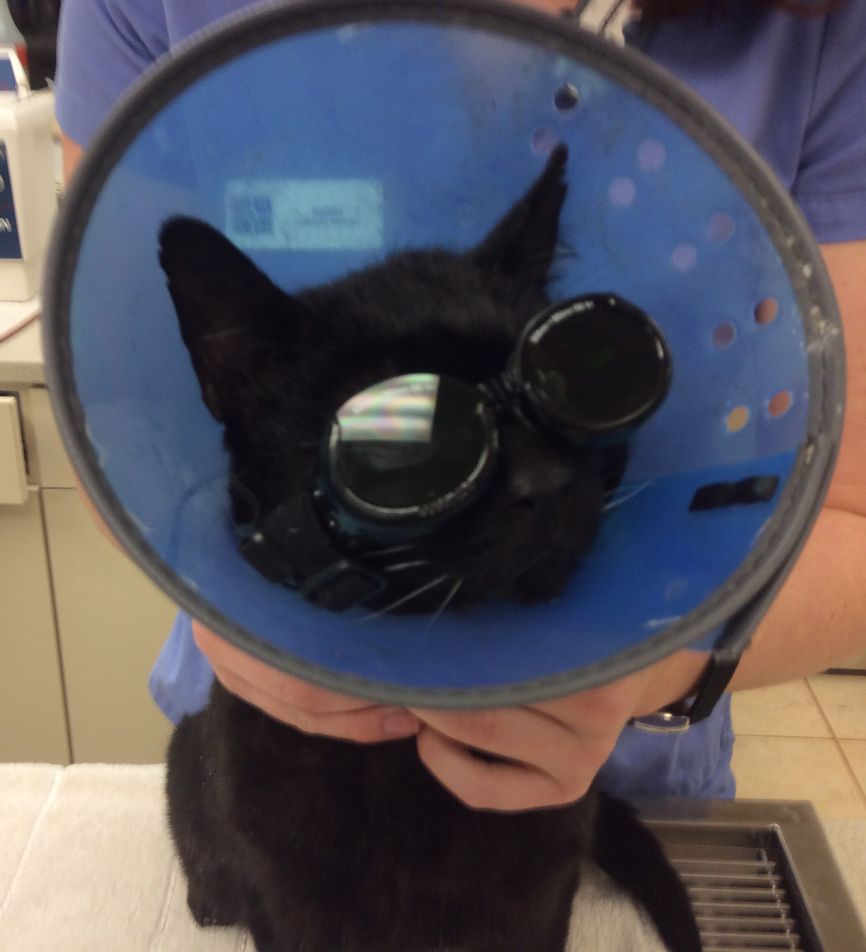 There are other tools we can use to shield the entire head of the patient itself, such as with the Comfy Cone® Elizabethan collars, made of dark, soft material that is comfortable for the patient to wear. The savvy laser operator may also have an assistant apply a series of distraction techniques for the patient during treatment sessions as well—simply petting the dog or cat, or offering food treats to keep the patient from investigating the laser treatment being administered.
Lastly, we also need to remember the required signage that needs to be prominently displayed when the laser is in use. Again, the manufacturer needs to provide this signage as it may slightly vary between different units. Each entryway leading in or out of the room where the laser is being used needs to have this sign as an added measure of safety.
It is important to remember that laser safe eye protection is not only our primary safety parameter, it is also one of the few and yet very specific guidelines set out by OSHA when it comes to the application of photobiomodulation therapy (PBMT). However, meeting this safety standard is easily achieved and should never be an issue in a clinical setting.
Physical Exam
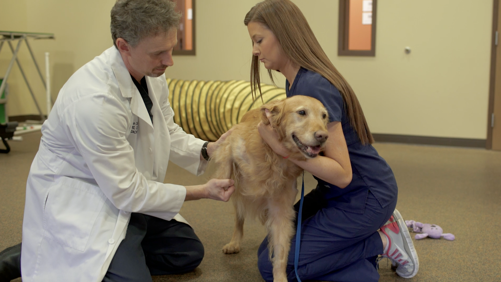 Performing a systematic physical examination and visual observation of the patient’s mobility will provide an initial assessment on where the potential issue(s) may exist. A thorough orthopedic exam allows for palpation of the limbs and joints while moving them through their range of motion to draw attention to changes within the tissue and help assess if the patient seems painful. Additionally, it is important to complete a neurologic evaluation and consider running additional diagnostic tests to look for any underlying disease processes that could affect treatment options.
Pain Evaluation
According to the Summary of 2015 AAHA/AAFP Pain Management Guidelines for Dogs and Cats, the most accurate method for evaluating pain is through observing any changes in the pet’s behavior. A pain score is considered the fourth vital sign after temperature, pulse and respiration.1 A patient’s pain should be evaluated in the clinic and by the pet owner to paint a full picture of how the pet is acting.
Pet Owner
Feedback from the owner is vital in understanding where, the length of time and severity of how the pet has been affected to determine the next steps for evaluating if arthritis is present. He or she should be asked a series of questions about any changes in behavior and given a pain scale form to fill out based on whether it is an acute or chronic problem. This will provide valuable insight into the pet’s everyday activities and how they vary.
Standing Evaluation
 Gathering data on how a patient is bearing weight while standing can provide objective numbers to determine which limb or limbs are effected. It has been shown that bathroom scales can be used as a reliable way to measure static weight bearing in canines2; therefore, a free-standing platform with built-in sensors, such as the Stance Analyzer, can visually show where a potential lameness exists. This sort of tool takes up minimal floor space and can be a cost-effective option in helping obtain a diagnosis, as well as measuring patient outcomes. Recording this sort of information can help guide treatment plans and provide owners with a better understanding of the source of their pets’ complications.
Gait Analysis
Objectively measuring a patient’s gait can be performed via force plate or a commercially available portable walkway. Force plate is considered the gold standard for evaluating gait by calculating the ground reactive forces of a cat or dog while standing, walking or jogging over the plate. The portable walkway is a mat that allows a pet to stand, walk or jog providing information on gait symmetry, stride length and the amount of pressure placed on each paw. These types of devices are expensive and take up a lot of floor space, thus they are not as common of an option for general practice.
Imaging
It is very important to obtain a picture of the joints suspected of having osteoarthritis. Radiographs are most commonly taken to assess the bony changes associated with OA. MRI and CT offer additional diagnostic capabilities, though less commonly used to assess OA, with MRI showing how the soft tissues are involved, while CT can offer better detail for the bony changes in more complex joints.
Arthrocentesis
Collecting a sample of the fluid in a swollen joint or joints suspected of having arthritis can help rule out what is causing the swelling or pain, especially in younger patients, or in those with a more complicated clinical history. The color, consistency and cellular make-up of the synovial fluid can provide clues as to what changes are occurring within the joint and why.3
References
1. Epstein, M. et. al. (2015). 2015 AAHA/AAFP Pain Management Guidelines for Dogs and Cats. JAAHA, 51(2), 67-84. doi: http://dx.doi.org/10.5326/JAAHA-MS-7331. 2. Hyytiäinen, H.K. et. al. (2012). Use of bathroom scales in measuring asymmetry of hindlimb static weight bearing in dogs with osteoarthritis. Veterinary and Comparative Orthopaedics and Traumatology, 25(5), 390-396. doi: https://doi.org/10.3415/VCOT-11-09-0135. 3. Degner, D. (2014, August). Arthrocentesis in Dogs. Clinician’s Brief. Retrieved from https://www.cliniciansbrief.com/article/arthrocentesis-dogs.

