HomeCommunityLaser Therapy Blog

Laser Therapy Blog

Learn tips about Class IV laser therapy and other health related topics on the Companion Therapy Lasers blog!  Check back weekly for updated posts.

8 Whys, Whens, and Hows for Deep Tissue Applicator (On Contact) Laser Therapy

Guest Blog by Jeff Smith, DVM, CCRP, Middletown Animal Hospital, Middletown, CA

8 whys whens and hows of deep tissue applicator laser therapy

1.  HOW:  The Deep Tissue Applicator (on contact) lenses are applied directly to the skin.  As such, the tissue is compressed (less distance), and blanched (fluid displaced—especially blood with hemoglobin).  Moreover, the hair coat is “parted” and displaced (diminishing unintended absorption at the surface).  Last, much less light is reflected from the skin when the lens is directly introducing the light into the tissue.  Altogether, a measurable 90% more photons reach deep tissues  than when applied with a non-contact handpiece.

2.  HOW:  Apply with moderate pressure, move the probe 1-3 inches per second, keep the probe perpendicular, move in a grid-like pattern, take the area through a range of motion, and treat the area from 360 degrees when possible.

3.  HOW:  3 Levels of Expertise:
One:  Deliver the therapeutic dose as described above.
Two:  Elicit a myofascial trigger point release with a pleasant massaging technique .
Three:  Determine diagnostic information based on the pets response (moving away from or into the pressure) to the application—use this to gauge the patient’s progress or determine areas of sensitivity.

4.  HOW:  Keep the heads clean and scratch free.  The heads can be cleaned inside an out with alcohol and a 3X3. Keep the red lens caps in place when the heads are not in use and keep one head on the handpiece at all times.

5.  WHY:  Dosing the tissue consistently and effectively is the key to maximum tissue response.  If 90% less of the intended dose is reaching the deeper target tissues, then one should expect much less effective response from those target tissues when using a non-contact head.

6.  WHEN:  Deep Tissue Applicators should be used whenever the target pathology is not visible to the eye.  In other words, any pathology that requires penetration of the energy through more that 5-10 mm of tissue:  Arthritis, IVDD, fractures, osteotomies, internal organs, deep tissue around the ear, deep tissue around the month, tendons, ligaments, and so on.  Occasionally animals may be uncomfortable enough that the initial treatment(s) need to be accomplished with a non-contact head until enough pain relief allows the use of the contact head.  Use the contact head(s) at 7-30 J/cm2.

7.  WHEN:  Non-contact heads can be used if you can see the damaged tissue:  Hot spots, superficial wounds, burns, minor incisions, external ear canal/pinna, exposed intraoperative organs, and so forth.  Use the non-contact head(s) at 3-4 J/cm2.

8.  WHEN:  Some protocols require both Contact and Non-contact heads (ears and mouth in particular).  This type of treatment effectively allows a much more thorough treatment from both the “inside” and the “outside” of the tissue.  A similar approach can be used with cases like a post-op TPLO:  Treat over the incision with a non-contact head/dose, then treat 270 degrees around the incision with the contact head/dose.
Posted in Featured News, Laser Therapy Blog | Leave a comment  

Read more

8 Ways to Treat Pain in Cats

Guest Blog by Jeff Smith, DVM, CCRP, Middletown Animal Hospital, Middletown, CA


8 ways to treat pain in cats

The new AAHA/AAFP Pain Management Guideline state that pain management should include pharmaceutical as well as non-pharmaceutical modalities.  MultiModal Pain Management is the new Standard of Care for painful patients
First, check laboratory values (CBC/Chem/UA), BP, and imaging.  This is not an exhaustive review of the subject, just a quick summary to act as a checklist of items to study and consider.
DJD is rarely evident on radiographs of cats.  Look for muscle atrophy, lack of jumping and lack of activity as the main indicators of joint pain.
1.  NSAIDs (COX Inhibitor):  Meloxicam is our drug of choice for safety, ease of use, and the ability to titrate or adjust the dose (0.02-0.03 mk/kg SID).  This is an off-label use in the US, but an approved use in Europe and Australia.
2.  Laser Therapy (decrease pain, decrease inflammation):  With contact head administration—3X per week for 4 weeks then 1X week for 1 month then once every 2-4 weeks.  Treat at 10-15 J/cm2 depending on severity.
3.  Weight Loss—Hill’s Metabolic Diet:  Weight loss is equally effective as NSAIDs for pain relief in arthritic patients.  This is often an overlooked but important consideration for geriatric cats.
4.  Adequan: Twice weekly for 6-8 doses (0.5 to 2.5 mg/lb IM or SQ).  Then every 4 months administer twice weekly for 4 doses—this protocol stimulates up regulation of the chondrocytes that will not occur with once monthly injections.  This is a much preferred and more effective joint therapy than oral supplements.  This the only disease modifying drug we have to use.
5.  Gabapentin (Ca Channel): Amantadine (NMDA), Amitriptyline (NE/Serotonin RUI), Bupirnex (Opiod).  All 4 agents block the pain pathway at different and unique locations, so they can be used in combination for added effect as needed. Look into the dosages and combined use of these meds as a more advanced extension of this discussion.
6.  Omega 3 Fatty Acids/Fish Oil (reduce Arachidonic Acid):  High EPA content and high quality.  WelActin is our neutriceutical of choice in this category.  We give a double dose (640 mg EPA/day/10# Cat) for the first 30 days and then reduce to a maintenance dose (320 mg EPA/day/10# Cat).
7.  Environmental Modification:  Ramps, steps, lower litter box barriers, rug runners—all help painful kitties get where they want to go with less discomfort.
8.  Physical Rehabilitation:  Yes!  Cats are candidates for rehab, too.  Underwater Treadmill, Massage, Passive Range of Motion, Strengthening Exercises, Core Exercises.  This is a rewarding and exciting aspect of veterinary medicine to develop.  Clients LOVE bringing their pets to rehab!
Consider advanced training such as the CCRP certification offered by the University of Tennessee or consult Darryl Millis’ Canine Rehabilitation Textbook.


Posted in Featured News, Laser Therapy Blog | Comments Off  

Read more

4 versatile applications for UWTM

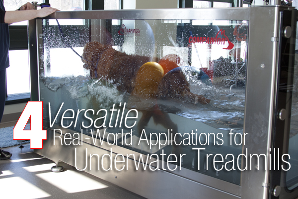
4 Versatile Real-World Applications for Underwater Treadmills
Guest Post by Erica Shoults, DVM

Are you considering adding an underwater treadmill to your clinic?  Learn about the top 4 clinical applications for the underwater treadmill!

Post Surgical Rehab
This is the number one reason why practices add an underwater treadmill to their clinics.  Patients recovering from post-operative TPLO surgery are one of the most common patients that benefit from the underwater treadmill.  The buoyancy of the water allows the dog to exercise in a controlled environment while minimizing weight bearing on the joints.  This helps him/her work on safely increasing their range of motion and lessens the loss of muscle mass that often occurs after surgery.  Other surgical patients, such as those who have had hip, elbow and back surgery, benefit greatly from aquatic therapy as well.

Weight Loss
According to a 2014 survey from the Association for Pet Obesity Prevention, an estimated 52.7% of dogs are overweight or obese.  Part of the issue is nutrition but the other contributing factor to canine obesity is lack of exercise.  The underwater treadmill allows an obese patient to exercise comfortably while the practitioner controls the speed and depth of the water.  The ability to control the exercise environment is especially important because the majority of these dogs also concurrently suffer from osteoarthritis in addition to obesity.  The warm soothing temperature of the water can also provide an additional therapeutic benefit by warming up muscles and joints.

Conditioning
The underwater treadmill can be an effective tool for exercising the canine athlete.  It provides the pet owner an option for keeping a dog in shape during the cold winter months.  Featuring speeds going up to 7 mph, and the flexibility to add difficulty by increasing or decreasing the height of the water, the underwater treadmill can facilitate generation of enhanced muscle mass and increase the cardiovascular fitness of the athlete.  Additional features, such as jets and incline, create added resistance to make the workout as challenging as possible.

Diagnostic
Another, and not so obvious use for the underwater treadmill, is that it can be used as a diagnostic tool in regards to gait and/or neurologic deficits.  Featuring tempered glass on all four sides of the chamber, the practitioner can walk around the unit and judge the patient’s gait at all angles.  Dogs will walk with a more exaggerated gait pattern than normal when they are in the water, this exaggeration can highlight an issue in a particular joint or area that might not have been observable while walking on land.  The gait ruler along the bottom of the glass on the underwater treadmill will also allow you to obtain the length of his/her stride to compare and contrast the dog’s progress from session to session.

Click here for more information about NEW Companion’s Underwater Treadmill.

Posted in Featured News, Laser Therapy Blog | Comments Off  

Read more