
Learn tips about Class IV laser therapy and other health related topics on the Companion Therapy Lasers blog! Check back weekly for updated posts.
With the growing area of regenerative medicine, it’s no surprise that there are dozens of systems available to process Platelet Rich Plasma (PRP). While the use of each system will result in a plasma product, the processing techniques, equipment and final sample differ quite drastically. In this installment of our Myth Buster series, we will take a closer look at the differences that one can expect to encounter when looking at PRP processing systems and how each of these components effects the PRP product.

Processing Techniques
There are three main methods by which Platelet Rich Plasma can be processed. These methods are:
1. Centrifugation – This is the most common technique available, which involves using a centrifuge to separate the cell layers in whole blood or to pass the blood components through a gel separator (which is in the device prior to insertion of blood) to isolate the platelets from other cell types.
 a. Spinning without gel separator – When blood is spun at a high rate of speed for a period of time, the cellular components will separate according to their density within the container/tube. These cell layers can be visualized upon removal of the device as seen in the below picture. The three distinct layers of blood include the plasma layer, the platelet buffy coat and the red blood cell layer. The plasma and buffy coat can be collected and injected or the suspension can be placed in a second container for a second spin. The second spin in the process enables further concentration of the platelets, which as we discussed in our previous posts, is important for delivering a therapeutic concentration of platelets (between 3-7-fold concentration compared to circulating blood). A sample that is obtained by a single spin process will have a lower concentration of platelets, thus possibly resulting in a sub-optimal therapy.
b. Spinning with gel separator – Gel separators provide a physical gradient for blood to pass through resulting with the final product at the top of the tubes. Gel separators are typically used for isolating either plasma or serum, but have been developed to allow smaller cell types to remain in the final product. Unfortunately, the majority of products available that use gel separation produce a low concentration of platelets due to the small volume of blood being used (typically 8- 10 mL) as well as the poor capture of platelets in the final product.
2. Flow Cytometry - This method involves a highly specialized piece of equipment that utilizes the absorption of light by a cell and further separates the cells according to the refracting light. This process relies on blood flowing in a single layer suspension through a complex network of tubes, which can be further complicated if the sample begins to clot during the process. Flow cytometry is typically automated, which provides great convenience to the user, but the equipment may require additional set up (prior to processing) and upkeep to ensure proper calibration for processing samples.
3. Gravity and Reverse Osmosis – This method involves utilizing a specially designed blood containment system with a built-in filter. The filter’s purpose is to collect the platelets while allowing the other cells to pass through. Although this method is attractive from a space and equipment perspective, performance of these systems has shown to result in an increase of neutrophils which can be detrimental when injected into a joint.
Volume of Blood
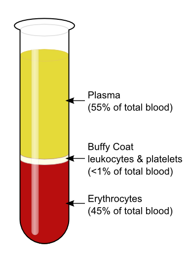 One of the biggest differences between PRP processing systems is the volume of blood that is required to obtain the PRP. Some systems require as low as 8 mL of whole blood, while other systems, such as the CRT system, require between 25-50 mL. What does this variation mean to you? If we look at typical blood composition, as depicted in the picture below, we will see that platelets make up less than 1% of the circulating blood. In order to have a therapeutic dose of platelets in the volume which is necessary for administration, a higher volume of blood is required for processing. For example, when using the CRT system, it is recommended to collect 50 mL of whole blood to produce 4-5 mL of PRP (with concentration of platelets between 3-7 fold). For systems that require the smaller whole blood volume with the same end product volume, the concentration of platelets will be below the recommended therapeutic threshold.
Sterility/ Exposure to Environment
When considering any product that will be administered intra-articularly or into areas of poor blood flow, it is imperative to ensure that the processing is done with aseptic technique. It is also important that the exposure of the sample to environmental factors is minimal to reduce the likelihood of contamination. The majority of systems available are considered to be closed systems in which the sample being processed has little to no contact with the outside environment. There are some processing systems available which utilize open tubes or require passing the blood through a needle into the containment device, which may introduce additional contaminants into the sample.
Time to Process- From Collection to Administration
Depending on the technique of isolating the platelets (centrifuge vs. flow cytometry vs. gravity filter), processing times vary greatly from just under 10 minutes to as much as 45 minutes! Some processing systems require activation of the platelets, which can take up to 45 minutes. Activating platelets, however, is not a necessary step, since the platelets will activate and release their stored growth factors once they are exposed to collagen in the joint/tissue. The Companion Regenerative Therapies System takes less than 15 minutes from blood collection to administration, making it a fast, in-house therapy that can be easily scheduled even in a busy hospital.
To learn more about Platelet Rich Plasma and how it works as a therapy, watch this short animation:
Stay tuned for our next blog post where we “bust” another regenerative medicine myth!
Guest Post by Matt Brunke, DVM, CVRPP, CVA, CVPP
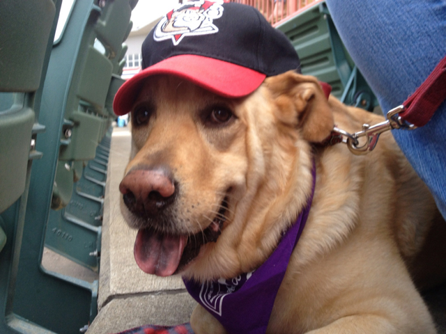
I am a big proponent of rescue groups. There are many “discarded” dogs and cats, most of which are absolutely wonderful pets. I have worked with Peppertree Rescue for many years and really appreciate their dedication to dogs with medical needs. Peppertree will find out about them and screen them, and then have them transported to the Albany area where they can be attended to. This was the case with Glinda, a sweet, 8-ish year old Golden Retriever cross.
 Upon physical exam, I found some areas of concern. While Glinda weighed in at a reasonable 74 pounds, her body condition score was abnormal. She had poor muscle mass in her back legs and was carrying more fat than she should. I also noted that she had decreased extension in both of her hip joints and was uncomfortable when I manipulated them. As I palpated her knees, I found chronic thickening in both the left and right knee with discomfort and mild instability. These findings are consistent with arthritic change in both the knees and hips.
Often times the hip arthritis is a consequence of hip dysplasia, while the arthritis and instability in her knees was consistent with chronic damage to her cruciate ligaments. I discussed this with the owners and we started with a conservative management program. I prescribed a course of therapy for her in the Rehabilitation Program’s Underwater Treadmill and an Adequan injection series for her. The buoyancy the underwater treadmill provides would allow Glinda to exercise and lose weight, while the Adequan injection series would be twice a week for 4 weeks, then one injection a month thereafter. This would help to improve her joint fluid in all of her joints, as well as reduce the damage to her cartilage.
Over the next 3 months, Glinda worked in the underwater treadmill twice a week, gradually increasing her time and distance with each session. Her comfort level improved and she lost 9 pounds. While she was moving well, I noted further instability in both of her knees. It was time for a sedated orthopedic exam and radiographs. Peppertree Rescue agreed to help with her costs and a short while later we had our answers.
Glinda had partially torn the cruciate ligament (ACL) in both of her knees. The right was not as stable as the left, and both had moderate arthritic change. She also had hip dysplasia, characterized by poor coverage of her femoral heads, and consequently had developed arthritis in her hips as well. The right knee needed stability. With hip arthritis in both legs and arthritis in both of her knees, the instability in the right needed surgical correction. My plan was to correct that with a procedure called a TPLO: TIbial Plateau Leveling Osteotomy. This would eliminate the need for the cruciate ligament and allow stability in the knee that would allow her to walk on the leg appropriately and slow the progression of arthritis forming in that knee. I was unsure if her left knee would need the same correction down the road.
Surgery was a success for Glinda. The TPLO gave her right knee the stability her body needed. She did very well in her post-operative rehab program, going through laser therapy treatments and underwater treadmill sessions. During her therapy she was always a smiling, happy patient. After 12 weeks of restricted activity, her tibia healed and I started to increase her overall activity. We would manage the arthritis in her knees and hips as an ongoing condition.

She has become an avid hiker with her owners. She goes for 30-45 minute walks every day and enjoys being outside. While her arthritis has progressed, her multimodal pain management regimen has kept her from having any further surgeries at this time.
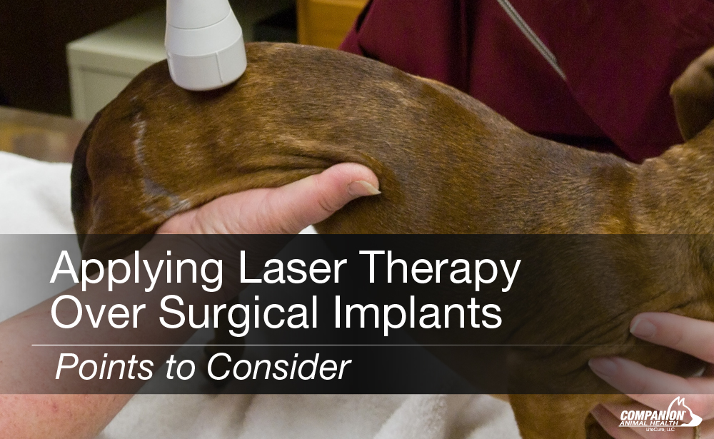
The application of laser therapy to sites containing surgical implants is perfectly safe. These implants will not heat up or be affected in any negative way when the treatment is carried out appropriately. Laser therapy is used to decrease postoperative pain while enabling the patient to experience a shorter convalescence period before returning to function.
Let’s consider what happens with tissue sites that have metal hardware. The laser light will be reflected from the implant and sent back through the tissues. Thus, if a bone surface has a metal implant over it, the tissues between the surface of the implant and the surface of the tissues will be exposed to more photons than the rest of the target site. To account for this, the laser operator simply needs to 1) increase their scanning speed when they are treating over an implant overlaying a bone and 2) treat 360° around the limb to ensure light penetrates to underlying fracture lines or osteotomy sites.
Target sites with cerclage wire and IM pins can certainly be treated as well. Because cerclage wire will cause minimal reflection, there is no need to increase scanning speed with these sites. The same is true for IM pins since they are located inside the bone itself, not on its surface.
Regardless of the specific case, the laser operator will constantly monitor the patient for any signs of discomfort and may possibly adjust the power settings down and/or increase scanning speed accordingly, as some very superficial implants (or very small patients with little soft tissue overlying the implant) may be uncomfortable.
The conscientious laser operator will be mindful when treating any patient with surgical implants, as these sites often develop arthritic changes as time goes on. In this manner, the presence of surgical implants actually predisposes patients for laser therapy in their later years.

