Signalment: Canine, 9yrs., F/S, Pit Bull, “Honey”.
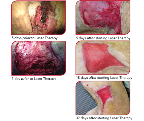
History: Patient wandered off and returned with skin wounds. Patient was initially anesthetized for wound debridement upon presentation. Surgical revisits were required to further remove devitalized tissues five and nine days after the initial procedure. Laser treatment started on day ten.
Diagnosis and Exam Findings: Large necrotizing cutaneous wound on the flank involving the fascia and superficial musculature.
Laser Treatment: 5J/cm2 delivered to target tissues, adjusting the total target dose as the treatment area reduced in size. The wound treatment protocol was selected and the patient was treated with an off contact technique.
Frequency of Treatments: Photobiomodulation therapy was initiated at day ten, as a daily treatment for ten days. Over the next two and a half months, the treatment frequency was reduced from every other day to twice weekly and then weekly treatment. In Total, the patient received about thirty treatments over an eighty day period.
Other Treatments Included: Standard of care including antibiotics, NSAID, and oral pain management was implemented, as well as daily wet to dry bandage changes for ten days.
Comments: Although laser therapy was not an initial part of the treatment plan, it quickly became apparent this large necrotizing wound would require the use of the modality. The patient quickly and consistently responded to photobiomodulation therapy until resolution.